Preventing heart disease linked to your dog’s teeth isn’t just about surface cleaning; it’s about stopping a systemic invasion that begins at the gumline.
- Bacteria from diseased gums constantly enter the bloodstream (bacteremia), forming infectious vegetations on heart valves.
- Anesthesia-free cleaning is purely cosmetic and dangerously ignores the fact that nearly 90% of periodontal disease occurs below the visible gumline.
Recommendation: For a dog with a heart murmur, a comprehensive, anesthetized dental assessment and treatment is not a ‘risk’ to be avoided—it’s an essential intervention to halt the progression of heart disease.
As a veterinary cardiologist, I often find myself in a delicate conversation with concerned owners. The discussion begins with the diagnosis of a heart murmur in their beloved, often small and aging, dog. Then, the conversation pivots to their pet’s mouth, and I see the confusion: “What do my dog’s bad teeth have to do with their heart?” This question is the critical starting point for understanding and preventing a serious condition called infective endocarditis. The common advice to simply “brush more” or offer dental chews is woefully inadequate once significant dental disease has taken hold, especially when a heart condition is already present.
The reality is that your dog’s mouth is not an isolated system. It is a gateway. When the gums are unhealthy, they lose their integrity as a protective barrier, allowing a constant shower of bacteria to enter the bloodstream. This article will not just tell you that dental disease is “bad.” Instead, we will connect the dots systemically, tracing the precise path from a broken oral barrier to a damaged heart valve. My goal is to reframe your perspective: managing your dog’s oral health is not a secondary chore but a primary strategy in managing their cardiovascular wellness.
We will explore how this bacterial migration happens, assess the true risks and necessities of dental procedures in a dog with a heart murmur, and differentiate confusing symptoms like coughing. We will also cover proactive nutritional strategies to support heart health and demystify the dangers of cosmetic, anesthesia-free procedures. This is a systemic approach to a systemic problem, designed to empower you to make the best decisions for your dog’s long-term health.
This guide provides a comprehensive overview, from the initial bacterial invasion to the clinical signs of complications. Follow along to understand the critical connections and the actionable steps you can take in partnership with your veterinarian.
Summary: How to Prevent Endocarditis by Maintaining Your Dog’s Oral Barrier?
- How Bacteria Travels From the Gumline to the Mitral Valve?
- Pulse Therapy: Should You Use Antibiotics Before Dental Cleaning?
- Grade 3 Murmur: Is Dental Surgery Too Risky or Essential?
- Coughing at Night: Is It Tracheal Collapse or Endocarditis?
- Can Omega-3 Fatty Acids Protect Heart Valves From Inflammation?
- Anesthesia-Free Cleaning: The Cosmetic Procedure That Hides Disease
- The Sodium Limit for Senior Dogs With Early Murmurs
- How to Recognize the Early Signs of Acute Organ Failure in Dogs?
How Bacteria Travels From the Gumline to the Mitral Valve?
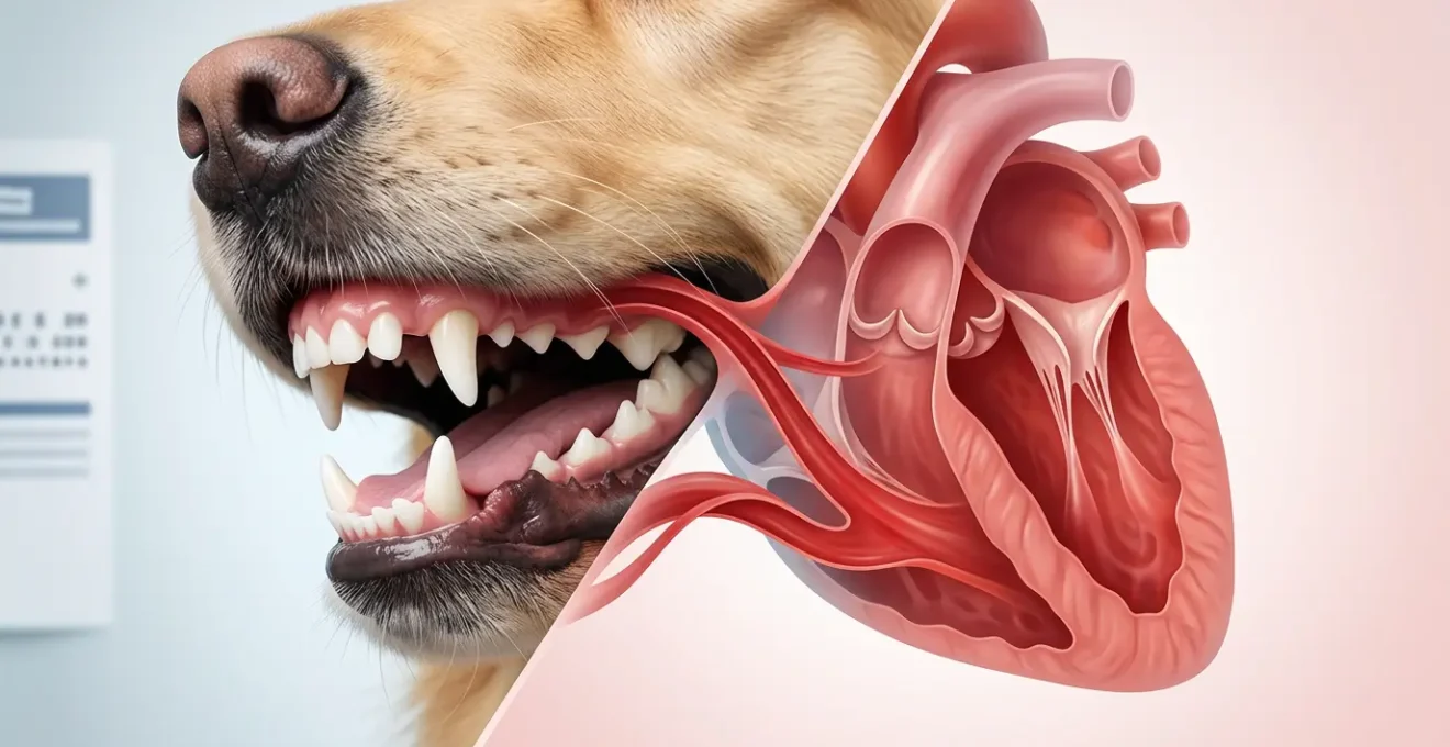
The journey from a diseased gum to a damaged heart valve is not instantaneous; it is a story of a compromised defense system. The critical structure is the oral barrier, specifically the thin layer of tissue called the sulcular epithelium, which lines the small pocket between the tooth and the gum. In a healthy mouth, this barrier is intact and robust. However, in the face of periodontal disease—a condition affecting an estimated 80-90% of dogs by age three—this barrier breaks down. Plaque, a biofilm of bacteria, accumulates at the gumline, triggering inflammation (gingivitis). This chronic inflammation makes the oral barrier weak and permeable.
This breakdown opens a direct superhighway for bacteria into the body. Every time your dog chews or even during routine daily activity, bacteria from these deep periodontal pockets are pushed into the rich network of capillaries in the gums. This event is called transient bacteremia—a shower of live bacteria entering the bloodstream. Once in circulation, these bacteria travel throughout the body. The heart, pumping the entirety of the body’s blood, is a primary target. Turbulent blood flow, often caused by a pre-existing condition like a leaky mitral valve (common in older, small-breed dogs), creates a perfect environment for these bacteria to attach to the valve leaflets.
Once attached, the bacteria colonize and form a “vegetation”—a clump of bacteria, inflammatory cells, and clotting factors. This is infective endocarditis. This vegetation not only physically damages the valve, making the leak worse, but it can also break off, sending septic emboli to other organs like the kidneys or brain. A case study of a Boston Terrier highlighted that dogs with severe periodontal disease have a six times higher risk of developing endocarditis, demonstrating a direct link from bacteremia secondary to dental disease to multi-organ septic embolization.
Pulse Therapy: Should You Use Antibiotics Before Dental Cleaning?
Given that we know dental disease creates a state of chronic bacteremia, the question of using antibiotics before a dental procedure is a logical one. The procedure itself, which involves scaling below the gumline and potentially extracting teeth, will inevitably release a significant bacterial load into the bloodstream. In fact, research shows that bacteremia occurs in roughly two-thirds of dogs after a dental prophylaxis, even when prophylactic antibiotics are used. This highlights that while antibiotics can reduce the bacterial load, they don’t eliminate the risk entirely.
The strategy of “pulse therapy” involves administering antibiotics for several days leading up to and following the dental procedure. The goal is to reduce the overall bacterial burden in the mouth *before* the main event, thereby lessening the intensity of the bacteremia that occurs during the cleaning. This is particularly considered for dogs with known cardiac risks or severe, purulent dental disease. The choice of antibiotic is critical and should be based on the types of bacteria commonly found in the canine mouth, such as anaerobes.
However, the veterinary community’s view on routine pre-emptive antibiotics is nuanced. Overuse of antibiotics contributes to resistance, a major global health concern. Furthermore, some research brings the direct link between a dental *procedure* and endocarditis into question. As a study by Glickman et al. noted:
Results did not provide any evidence of an association between bacterial endocarditis in dogs and either dental or oral surgical procedures.
– Glickman et al., PubMed Study on Association of periodontal disease and endocarditis
This does not mean the link is non-existent. It suggests the primary culprit is the chronic, daily bacteremia from the underlying disease, not necessarily the single, acute event of a cleaning. Therefore, the decision to use pulse therapy is a risk-benefit analysis performed by your veterinarian, weighing your dog’s specific cardiac status, the severity of their dental disease, and the principles of antibiotic stewardship.
Grade 3 Murmur: Is Dental Surgery Too Risky or Essential?
This is the most common and pressing fear I hear from owners: “Is the anesthesia for a dental cleaning too dangerous for my dog with a heart murmur?” It is a valid concern, as anesthesia does require the cardiovascular system to adapt. However, the question must be reframed: what is the risk of *not* performing the dental procedure? As we’ve established, severe periodontal disease is a constant source of systemic inflammation and infection. It is not a passive or stable condition; it is an active disease process that puts a continuous strain on the heart and other organs.
Data clearly shows the risk is with the disease itself. A study comparing dogs with and without advanced periodontal disease found that the prevalence of endocarditis was 0.15% in dogs with Stage 3 periodontal disease, compared to just 0.01% in dogs without. This represents a more than ten-fold increase in risk attributable to the dental disease itself. Therefore, leaving severe dental disease untreated in a dog with a heart murmur is not the “safe” option; it is a decision to allow a major risk factor for cardiac decline to continue unchecked.
The modern approach is not to avoid anesthesia, but to make it as safe as possible. For a dog with a Grade 3 murmur, this means a thorough pre-anesthetic workup and a customized anesthetic protocol. The goal is to stabilize the patient, understand their specific cardiac function, and tailor the drugs and monitoring to support them through the procedure. This is where a partnership between your general practitioner, and potentially a veterinary cardiologist and anesthesiologist, becomes vital.
Your Vet’s Pre-Anesthetic Cardiac Action Plan
- Referral and Imaging: A referral to a veterinary cardiologist for an echocardiogram (heart ultrasound) is often the first step to precisely evaluate heart structure and function before the dental procedure.
- Staging the Disease: The veterinarian will assess the ACVIM (American College of Veterinary Internal Medicine) stage of heart disease, classifying it from A (at risk) to D (advanced heart failure).
- Evaluating Dental Severity: The severity of the dental disease itself is graded on a scale of 1-4 to understand the extent of the necessary intervention.
- Tailoring Anesthesia: A customized anesthetic plan is created using cardiac-safe drug protocols that maintain blood pressure and cardiovascular stability.
- Multimodal Monitoring: During the procedure, the patient is intensely monitored using tools like ECG (electrocardiogram), direct arterial blood pressure, and capnography (CO2 monitoring).
Coughing at Night: Is It Tracheal Collapse or Endocarditis?
A nighttime cough in a small, older dog is a classic and alarming symptom that sends many owners to the clinic. The two most common culprits are often tracheal collapse and congestive heart failure. However, a cough can also be a sign of endocarditis, and differentiating them is crucial. Tracheal collapse, a structural weakening of the windpipe common in small breeds, typically produces a very distinctive, dry, “goose honk” cough, often triggered by excitement or pressure on the neck (like from a collar). The cough from heart disease is usually softer, wetter, and can be a sign of fluid accumulating in the lungs (pulmonary edema).
Endocarditis can present with a cough, but it is often accompanied by other, more systemic signs that are not typical of uncomplicated tracheal collapse. These include a persistent or intermittent fever, lethargy, loss of appetite, and sometimes shifting leg lameness if septic emboli lodge in a joint. A key diagnostic clue for endocarditis is the sudden appearance of a new, loud heart murmur or a dramatic change in an existing one. Because endocarditis can cause a wide array of symptoms depending on which organs are affected by septic emboli, its presentation can be highly variable.
Diagnosing the cause of the cough requires a thorough veterinary examination, including chest X-rays and often an echocardiogram. The following table provides a simplified comparison of key differentiating features:
| Symptom | Tracheal Collapse | Endocarditis |
|---|---|---|
| Cough Type | Goose honk cough | Soft, wet cough |
| Associated Signs | Tracheal sensitivity | Fever, lethargy, new murmur |
| Diagnostic Finding | Collapsed trachea on X-ray | Vegetative lesions on echo |
| Primary Cause | Structural weakness | Bacterial infection |
While this table, based on information from the Merck Veterinary Manual, offers a guide, only a veterinarian can make a definitive diagnosis. It is critical not to dismiss a new cough, especially in a dog with known dental and heart disease.
Can Omega-3 Fatty Acids Protect Heart Valves From Inflammation?
While addressing the source of infection through dental care is paramount, we can also use nutrition to support the cardiovascular system and mitigate the inflammatory damage. One of the most powerful tools in our nutritional arsenal is Omega-3 fatty acids, specifically EPA (eicosapentaenoic acid) and DHA (docosahexaenoic acid). These fatty acids, found abundantly in marine sources like fish oil, have potent anti-inflammatory properties. They work by altering the production of inflammatory mediators in the body, shifting the balance towards resolution rather than perpetuation of inflammation.
In the context of periodontal and heart disease, this is doubly beneficial. Omega-3s can help reduce the chronic inflammation in the gums, supporting the integrity of the oral barrier. Systemically, their anti-inflammatory effects may help protect the heart valves and heart muscle (myocardium) from the damaging inflammatory cascade triggered by bacteremia. While Omega-3s cannot prevent bacteria from sticking to a valve, they can help modulate the body’s overzealous inflammatory response that contributes to valve damage and scarring.
Beyond Omega-3s, several other supplements can play a supportive role in a cardiac-focused nutritional plan. These work together to support heart muscle function, provide antioxidant protection, and ensure the energy-producing mitochondria within heart cells are working optimally. A comprehensive plan should always be discussed with your veterinarian, as dosages and combinations will vary based on your dog’s specific condition.
Here is a protocol of nutritional supplements often considered for cardiac health:
- Choose marine-based Omega-3 sources (fish oil, krill oil) for their direct EPA and DHA content.
- Look for third-party tested products to ensure purity from heavy metals and potency.
- Consider Coenzyme Q10 (CoQ10) supplementation to support mitochondrial energy production in the hard-working heart muscle.
- Add antioxidants like Vitamin E to help combat the oxidative stress associated with chronic inflammation.
- Include amino acids like L-carnitine and Taurine, which are crucial for normal myocardial function.
Anesthesia-Free Cleaning: The Cosmetic Procedure That Hides Disease
The concept of “anesthesia-free dental cleaning” is appealing to many owners, especially those with pets they perceive as high-risk for anesthesia. These services promise a clean, white smile without the perceived dangers and cost of a veterinary procedure. However, from a medical and systemic health perspective, this practice is not just ineffective; it is actively dangerous because it masks the true extent of the disease.
The core of the problem lies in what you can’t see. Research confirms that the vast majority of periodontal disease—up to 90% of it—occurs below the gumline (subgingivally). Anesthesia-free cleaning, performed on an awake patient, can only scrape tartar from the visible surface of the tooth (the crown). It is impossible to safely and effectively clean the deep pockets under the gums where the most dangerous bacteria thrive. It also does not allow for dental X-rays, which are essential for evaluating the tooth roots and bone that make up over half the tooth structure.
This creates a false sense of security. The teeth may look white, but the engine of infection and inflammation beneath the gums continues to rage, seeding bacteria into the bloodstream and driving the very processes that lead to endocarditis. The procedure is purely cosmetic, like painting over a rusty car frame. The appearance is improved, but the underlying structural decay continues unabated. As veterinary experts at Today’s Veterinary Practice point out, the problem rebuilds itself quickly:
Plaque forms on tooth surfaces within 24 hours after complete cleaning.
– Today’s Veterinary Practice, Periodontal Disease: Utilizing Current Information
This rapid reformation of plaque underscores why the only effective treatment is a complete cleaning above and *below* the gumline, followed by daily at-home care. Anesthesia-free cleaning accomplishes neither of these and delays the essential care your pet needs.
The Sodium Limit for Senior Dogs With Early Murmurs
For a dog that already has a heart murmur, indicating some level of valve disease, managing the heart’s workload becomes a crucial part of their long-term care. One of the most direct ways to influence this is by controlling dietary sodium. High levels of sodium cause the body to retain water, which increases the total blood volume. For a heart with a leaky valve, this extra volume means it has to work significantly harder with every beat to pump blood, accelerating wear-and-tear and pushing the heart closer to failure.
The connection between advanced periodontal disease and congestive heart failure (CHF) is underscored by the chronic inflammatory burden. Studies have shown that periodontal disease is associated with cardiovascular conditions like cardiomyopathy and endocarditis, contributing to outcomes like CHF through systemic inflammation. Managing sodium is a cornerstone of managing an already-compromised heart.
Sodium restriction is not about eliminating it entirely, but about carefully controlling it according to the stage of heart disease. The ACVIM provides clear guidelines for veterinarians to follow. For a dog in the early stages (Stage B1: a murmur is present, but the heart is not yet enlarged), a mild restriction is often recommended as a proactive measure. As the disease progresses and the heart begins to enlarge (Stage B2) or enter failure (Stage C), the sodium restriction becomes progressively stricter.
It’s vital to remember that sodium hides in many places beyond the main diet, especially in treats. Dental chews, jerky, and table scraps can be loaded with sodium and can easily sabotage a carefully planned cardiac diet. Reading labels and discussing all treats with your veterinarian is essential.
- Stage B1 (murmur, no enlargement): A target of less than 100 mg of sodium per 100 kcal of food.
- Stage B2 (murmur with enlargement): The target tightens to less than 90 mg of sodium per 100 kcal.
- Stage C (congestive heart failure): A stricter limit of less than 80 mg of sodium per 100 kcal is often implemented.
- Hidden Sodium Audit: Always review all treats, dental chews, and table scraps for hidden sodium sources.
Key Takeaways
- The oral barrier (gum tissue) is the primary entry point for bacteria that can travel to and infect a dog’s heart valves, causing endocarditis.
- For dogs with stable heart murmurs, the systemic health risk of untreated, severe dental disease significantly outweighs the managed risk of a modern, monitored anesthetic procedure.
- Anesthesia-free dental scaling is a purely cosmetic procedure that fails to address the subgingival disease driving systemic inflammation and gives a false sense of security.
How to Recognize the Early Signs of Acute Organ Failure in Dogs?
The ultimate and most devastating consequence of untreated infective endocarditis is not just worsening heart disease, but acute organ failure. This occurs when the septic vegetations on the heart valves break apart, sending showers of bacteria-laden clots (septic emboli) throughout the body. Where these emboli lodge determines the clinical signs. If they go to the kidneys, it can cause acute kidney failure. If they go to the brain, it can cause seizures or stroke-like signs. If they lodge in the spleen or liver, it can lead to abscesses and organ dysfunction.
Recognizing these signs requires vigilance. The symptoms are often non-specific and can mimic other diseases, which makes diagnosis challenging. A dog may present with a sudden onset of severe lethargy, collapse, a high fever that doesn’t respond to typical treatments, or pain in their abdomen or limbs. Shifting leg lameness can occur if emboli lodge in the blood vessels of a limb or a joint. The most common bacteria isolated in these cases include species like Streptococcus, Staphylococcus, and E. coli, with Bartonella being a recognized cause of aortic valve endocarditis specifically.
The goal of everything discussed in this article—from maintaining the oral barrier to performing safe dental procedures—is to prevent the disease from ever reaching this critical stage. Preventing the initial bacterial invasion is infinitely better than trying to manage a systemic septic crisis. As a dog owner, your role is to be an astute observer. Any sudden, unexplained change in your dog’s health, especially if they have known dental and cardiac disease, warrants an immediate veterinary visit. Early intervention is the only chance to combat this aggressive complication.
Frequently asked questions on How to Prevent Endocarditis by Maintaining Your Dog’s Oral Barrier?
What are the signs of septic shock from dental disease?
The clinical signs of infectious endocarditis, which can lead to septic shock, frequently include a chronic intermittent or continuous fever, shifting leg lameness, weight loss, and profound lethargy.
How can I differentiate acute heart failure from other conditions?
Acute regurgitation (leakage) of the mitral or aortic valve due to endocarditis typically results in left-sided congestive heart failure. This manifests as pulmonary edema (fluid in the lungs), causing rapid breathing (tachypnea), difficulty breathing (dyspnea), and a soft cough.
What laboratory changes indicate organ involvement?
Bloodwork can reveal organ damage from septic emboli. A Complete Blood Count (CBC) often shows a high white blood cell count (neutrophilic leukocytosis) and anemia. Serum chemistry analysis may show increased liver enzymes or markers of kidney damage (BUN and creatinine), while substantial protein in the urine can indicate kidney damage from immune-complex glomerulonephritis.
Your partnership with your veterinarian is the most powerful tool in this fight. Schedule a consultation to discuss your dog’s specific oral and cardiovascular health, and work together to create a proactive, long-term plan that addresses the root cause of the problem, ensuring a longer, healthier life for your companion.